Bones Of Female Back - Female Backbone Hd Stock Images Shutterstock / During the second stage of labour, a combination of bones including your sacrum actually move backwards and in doing so, increases the diameter of your pelvis.
Bones Of Female Back - Female Backbone Hd Stock Images Shutterstock / During the second stage of labour, a combination of bones including your sacrum actually move backwards and in doing so, increases the diameter of your pelvis.. Pain in your upper or lower back, groin, buttocks, and thighs 7 The test can identify osteoporosis, determine your risk for fractures (broken bones), and measure your response to osteoporosis treatment. The vertebral column of the lower back includes the five lumbar vertebrae, the sacrum, and the coccyx. Gazzaniga notes that 1 in 2 women and 1 in 4 men will have a fracture over their lifetime due to osteoporosis, and that in the first five years after menopause, a woman can lose up to 20 percent. If you want to try pilates, look for classes that are specifically for women with bone health issues, or talk to the instructor of the class to understand how you can modify positions to protect your bones.
Powerful muscles that move the head and arms attach to these bones as well. The bones of the pelvis and lower back work together to support the body's weight, anchor the abdominal and hip muscles, and protect the delicate vital organs of the vertebral and abdominopelvic cavities. It runs down the centre of the body. This is what is known as opening of. It is the surface of the body opposite from the chest and the abdomen.the vertebral column runs the length of the back and creates a central area of recession.

It runs down the centre of the body.
This protects the spinal cord inside. Women also may lose bone mass during breastfeeding because they're producing less estrogen, which is the hormone that protects bones. Rare causes of lower back pain 5. The vertebral column of the lower back includes the five lumbar vertebrae, the sacrum, and the coccyx. The human back, also called the dorsum, is the large posterior area of the human body, rising from the top of the buttocks to the back of the neck. It represents a vestigial tail, hence the common term tailbone. Pain in your upper or lower back, groin, buttocks, and thighs 7 Spinal osteoarthritis causes a breakdown of the fibrous cartilage in the facet joints. What is a bone density test? There are two hip bones, one on the left side of the body and the other on the right. The spine is composed of 33 bones called vertebrae, which stack together to form the spinal canal. The coccyx is a triangular arrangement of bone that makes up the very bottom portion of the spine below the sacrum. This relieves pressure on spinal nerves and can ease pain or weakness, but the procedure can make.
Race and ethnicity worldwide, northern europeans and caucasians have the greatest risk of fracture due to osteoporosis. Rare causes of lower back pain 5. Pain in your upper or lower back, groin, buttocks, and thighs 7 Powerful muscles that move the head and arms attach to these bones as well. Related posts of anatomy of the back organs human body full parts inside.

The breadth of the back is created by the shoulders at the top and the pelvis at the bottom.
It is the surface of the body opposite from the chest and the abdomen.the vertebral column runs the length of the back and creates a central area of recession. Women also may lose bone mass during breastfeeding because they're producing less estrogen, which is the hormone that protects bones. The human back, also called the dorsum, is the large posterior area of the human body, rising from the top of the buttocks to the back of the neck. Gazzaniga notes that 1 in 2 women and 1 in 4 men will have a fracture over their lifetime due to osteoporosis, and that in the first five years after menopause, a woman can lose up to 20 percent. Spinal osteoarthritis causes a breakdown of the fibrous cartilage in the facet joints. The red lines point individual bones and the names are writen in singular, the blue lines conect to group of bones and are in plural form. Osteoarthritis of the lower back can cause: The breadth of the back is created by the shoulders at the top and the pelvis at the bottom. In this procedure, a surgeon removes parts of the bone, bone spurs, or ligaments in your back. The bones of the chest and upper back combine to form the strong, protective rib cage around the vital thoracic organs such as the heart and lungs. Women over the age of 50 have the greatest risk for developing the bone disease. The bones that make up your spine (vertebrae) can weaken to the point of crumpling, which can result in back pain, lost height and a hunched forward posture. Rare causes of lower back pain 5.
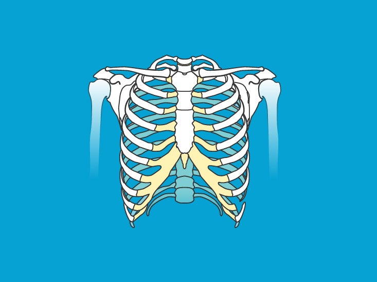
Everything else that hangs from this, like the arms, legs, shoulders, and hips, is called the appendicular skeleton. This relieves pressure on spinal nerves and can ease pain or weakness, but the procedure can make. During the second stage of labour, a combination of bones including your sacrum actually move backwards and in doing so, increases the diameter of your pelvis. It runs down the centre of the body. The rib cage also anchors the bones of the head, neck, shoulders, and arms to the trunk of the body.
Together, they form the part of the pelvis called the pelvic girdle.
It represents a vestigial tail, hence the common term tailbone. Race and ethnicity worldwide, northern europeans and caucasians have the greatest risk of fracture due to osteoporosis. Osteoarthritis of the lower back can cause: Diagram of a human female skeleton, back view. It is most common in postmenopausal women. Prevention good nutrition and regular exercise are essential for keeping your bones healthy throughout your life. So what are some good ways to build healthy bones? Bones make up about 14 percent of our body weight. The breadth of the back is created by the shoulders at the top and the pelvis at the bottom. The axial skeleton is made up of the skull, backbone, breastbone, and ribs. The rib cage also anchors the bones of the head, neck, shoulders, and arms to the trunk of the body. Pelvic floor female female pelvis bone pelvis medical anatomical illustration pelvic posterior pelvic girdle pelvic pelvis anatomy sacroiliac joint pelvic floor pelvis. The achilles tendon , located to the back and center of the ankle, above the heel, is injured about three times as often in men, miller says, although achilles tears are growing more common in women.


Komentar
Posting Komentar